Table of Contents
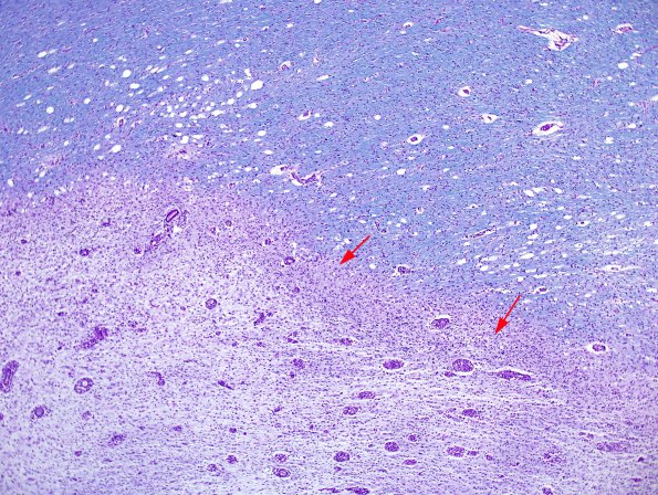
Washington University Experience | MYELIN (IMMUNE-MEDIATED) | MS - Secondary Progressive (SPMS), fulminant | 1E2 MS, Secondary Progressive, fulminant (Case 1) CC2 LFB-PAS 8 copy
1E2-4 These variable magnification images of myelin stains show an area of demyelination (lower left region, #1E2), the surrounding vacuolated near-normal white matter (right upper region, #1E2) and a rim of pink stained border between them (arrows) which represents a site of active demyelination. This lesion is shown at progressively higher magnifications in subsequent images. (LFB-PAS)

