Table of Contents
Washington University Experience | MYELIN (IMMUNE-MEDIATED) | NMO (Neuromyelitis Optica) | NMO Spectrum Disorder | 1A4 NMO (Case 1) MRI 2 T1 W 3 - Copy
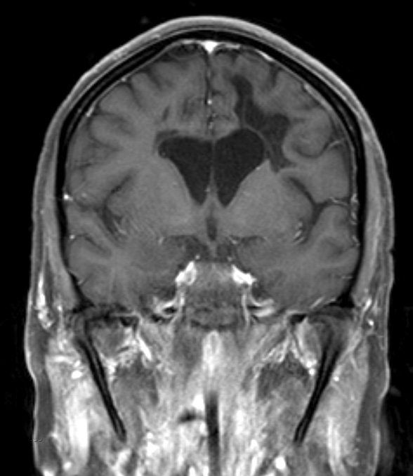
Coronal T1-weighted image with contrast shows marked periventricular tissue loss, extending into the subcortical digitate white matter and patchy hypointense lesions involving white matter. Notice the dilatation and blunting of the lateral ventricles.

