Table of Contents
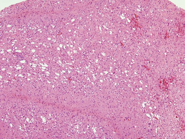
Washington University Experience | MYELIN (IMMUNE-MEDIATED) | Sentinel & Steroid Rx Lesions | 4B3 Lymphoma, B cell & sentinel (Case 4) demyelinated area H&E 3
4B3-5 Although the white matter is vacuolated with scattered areas of pallor and astrocytosis, this is not the typical appearance of demyelination which is usually more pale (myelin stains pink on H&E) with fewer empty vacuoles. The axonal swellings in image 4B5 (arrows) are also present in larger numbers than typical MS (H&E)

