Table of Contents
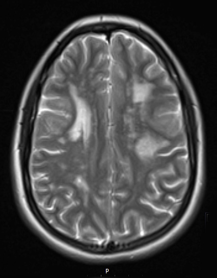
Washington University Experience | MYELIN (NON-IMMUNE MEDIATED) | CADASIL | 11A4 CADASIL (Case 11) T2 - Copy
Lesions are hyperintense in this T2-weighted scan without contrast. ---- MRI Comment: Some of these MRI lesions were ovoid, well demarcated, perpendicular periventricular lesions, as well as a corpus callosum genu lesion. These lesions have an appearance which could be seen with demyelinating disease but are atypical considering the degree of diffusion restriction, T1 hypointensity without contrast enhancement; this MRI appearance was thought more typical of ischemic disease. There were older areas of encephalomalacia were likely prior subclinical ischemic events. The clinical impression was that of an inherited arteriopathy, most likely CADASIL.

