Table of Contents
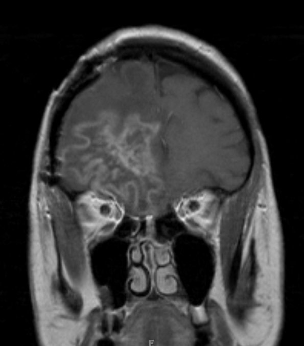
Washington University Experience | MYELIN (NON-IMMUNE MEDIATED) | Delayed Radionecrosis | 3A3 Radiation Necrosis (Case 3) T1 with contrast 2 - Copy
A coronal section of T1-weighted contrast administered MRI image shows a serpiginous border of hyperintensity as well as a surrounding rim of hypointensity.

