Table of Contents
Washington University Experience | MYELIN (NON-IMMUNE MEDIATED) | Fat Embolism | 1D5 Embolism, fat (Case 1) ORO 12 copy
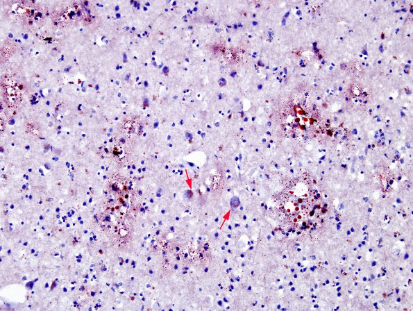
Notice that this image (adjacent to those previously shown) reveals microvascular fat droplets in the ORO stained image of the cerebral cortical gray matter. The aggregates of lipopigment within small neurons are also stained (arrows). Gray matter infarction and hemorrhages may not appear in size and numbers comparable to white matter pathology. This dichotomy may reflect protection by the anastomosing microvasculature which is 3-fold denser in cerebral cortex than in subcortical white matter (ORO).

