Table of Contents
Washington University Experience | MYELIN (NON-IMMUNE MEDIATED) | Neuroaxonal Leukodystrophy | 6A Neuroaxonal Leukodystrophy (Case 6)
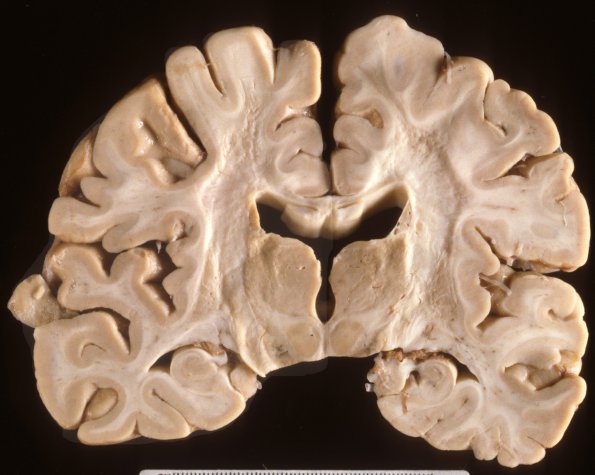
The brain weighed 1300 grams in the fixed state. On cut sections the cortical ribbon showed mildly atrophic changes with no evidence of infarcts or focal defects. The white matter revealed loss and granular gray-white discoloration of subcortical white matter into the internal capsules. The ventricles were enlarged and blunted. Bilateral thalamic discoloration and softening was not accompanied by caudate/putamen changes. The cut surfaces of the cerebellum showed a normal folial pattern. Serial horizontal sections of the brainstem reveal no gross pathology, specifically, there is no discoloration of the white matter. ---- This coronal section shows ventricular dilatation, loss of callosal white matter and mildly discolored, friable and shrunken white matter.

