Table of Contents
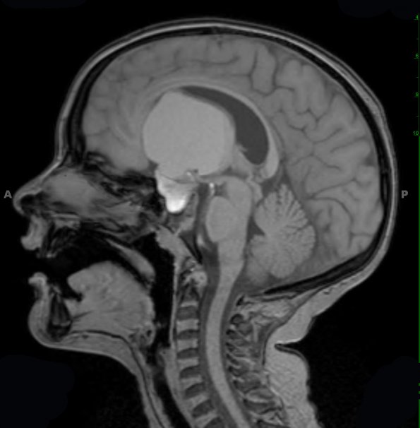
Washington University Experience | NEOPLASM (SELLAR) | Craniopharyngioma, adamantinomatous | 11A1 Craniopharyngioma (Case 11) T1 2 - Copy
Case 11 History ---- The patient is a seven-year-old boy with short stature and a two-week history of increasing headaches. MRI in February 2011 showed a 5.6 x 4.7 x 4.9 cm mass. Clinical impression: Craniopharyngioma. Operative procedure: Craniotomy for resection. ---- 11A1-4 Multiple MRI scans show a peripherally rim-enhancing, cystic sellar/suprasellar mass with a proteinaceous/hemorrhagic fluid level, centered in the sella/suprasellar region, with expansion of the sella. This T1-weighted scan is shown in sagittal (11A1) and axial (11A2) scans demonstrating the size and effect the mass has on the ventricular systems.

