Table of Contents
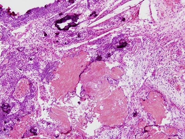
Washington University Experience | NEOPLASM (SELLAR) | Craniopharyngioma, adamantinomatous | 11B Craniopharyngioma (Case 11) H&E 1
Histological sections show a cystic neoplasm. Much of the cyst interior is lined by a relatively inconspicuous, thin, keratinizing squamous epithelium. In some histologically 'classic' areas, however, the epithelium shows a dense palisading basal layer, above which multiple poorly defined strata of elongate oval hyperchromatic nuclei reside, decorated by 'pearls' of so-called 'wet keratin' (anucleate squamous cells). Focally, this epithelium shows coarse vacuolation, leaving a thin meshwork of cell processes, or 'stellate reticulum.' Elsewhere, the inner surface is lined by reactive tissue resulting from a proliferant fibrohistiocytic response to the cyst contents. Within the cyst is an abundance of 'wet keratin', necrotic, grumous debris, abundant cholesterol clefts, dystrophic calcification, hemosiderin deposits, and granulomatous inflammation. In some areas, the cyst wall is formed by a thick layer of relatively hypocellular collagenous connective tissue; elsewhere, this layer is not observed. External to this collagenous tissue, the outer rim of the tumor shows a hypodense, richly vascular tissue. Parts of the outer surface of specimen C additionally show a thin layer of adherent, mildly gliotic brain tissue (without evidence of piloid gliosis).

