Table of Contents
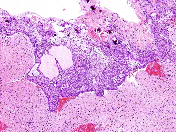
Washington University Experience | NEOPLASM (SELLAR) | Craniopharyngioma, adamantinomatous | 12A1 Craniopharyngioma (Case 12) 9
Case 12 History The patient is a seven year old boy with a complex, multilobulated, partially calcified supracellular mass causing obstructive hydrocephalus. Prolactin and cortisol are mildly elevated; FSH, TH and LH are within normal limits. Clinical diagnosis: Craniopharyngioma. Operative procedure: Craniotomy for tumor resection. 12A1-5 Sections of the suprasellar mass show a tripartite tumor composed of squamous islands with palisading peripheral basaloid cells, keratin pearls, calcifications and edematous interstitial tissue (the so-called "stellate reticulum"). The histologic relationship to the tumor and surrounding brain is well seen and characterized by astrocytosis and Rosenthal fibers.

