Table of Contents
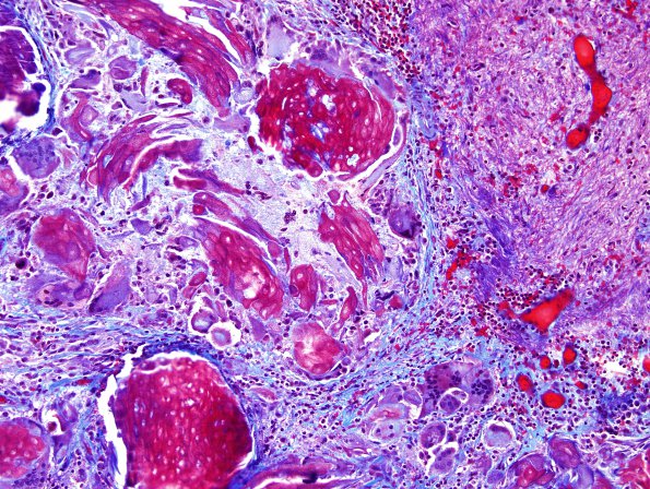
Washington University Experience | NEOPLASM (SELLAR) | Craniopharyngioma, adamantinomatous | 14C1 Cranio SP resection, no live tumor (Case 14) Trichrome 3.jpg
14C1,2 A trichrome stain highlights focal fibrotic tissues as well as adjacent hyalinized blood vessels, again, suggestive of a chronic process. The trichrome stain also incidentally highlights the wet keratin a magenta color. ---- Other stains (not shown): A CD45 immunostain highlights the numerous lymphocytic cells scattered throughout the tissues. A beta catenin immunostain is positive only in a cytoplasmic distribution of the glial tissues and blood vessels. Of note, mutational analysis for the BRAF V600E was attempted, but was not performed due to the absence of obvious tumor.

