Table of Contents
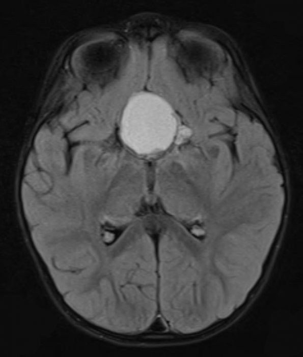
Washington University Experience | NEOPLASM (SELLAR) | Craniopharyngioma, adamantinomatous | 16A1 Craniopharyngioma (Case 16) FLAIR - Copy - Copy
Case 16 History ---- The patient is a 3 year old girl with a 6 month history of eye deviation and progressively worsening vision. She was found to have bilateral optic nerve atrophy. MRI demonstrated a large extra-axial, predominately cystic, partially solid, rim-enhancing sellar mass with focal calcification and peripheral nodular areas of enhancement. It also had suprasellar extension that resulted in compression of the optic nerves and chiasm. Operative procedure: Right craniotomy for tumor excision. ---- 16A1-6 MRI: 16A1 Hyperintensity is shown in this FLAIR image.

