Table of Contents
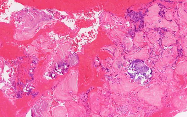
Washington University Experience | NEOPLASM (SELLAR) | Craniopharyngioma, adamantinomatous | 16B Craniopharyngioma (Case 16) H&E 10X
Sections show a proliferation of cells with bipolar nuclei and spindle cell features in a background of myxoid material but also demonstrates a microscopic focus of wet keratin and a fragment of palisaded epithelium. The portions of the mass submitted entirely for permanent sections show fragments of wet keratin, palisaded epithelium, and occasional small foci of stellate reticulum as well as blood and calcifications. In addition there are fragments of reactive astrocytic proliferation.

