Table of Contents
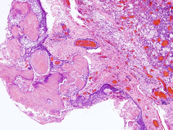
Washington University Experience | NEOPLASM (SELLAR) | Craniopharyngioma, adamantinomatous | 17A1 Craniopharyngioma (Case 17) H&E 4
Case 17 History ---- The patient is a 27 year old woman who presented with galactorrhea and amenorrhea. MRI shows a 2.6 x.2.5 x 2.6 cm, solid and cystic, partially calcified, avidly enhancing suprasellar mass. Clinical/radiological diagnosis: Craniopharyngioma. Operative procedure: Right craniotomy. ---- 17A1-6 Histological sections of the "suprasellar tumor" show multiple fragments of a partially cystic neoplasm, that exhibits an epithelial component with basal nuclear palisading and rarified areas ('stellate reticulum'); 'wet' keratin; and dystrophic calcifications. In addition, there is multinucleate giant cells reactive to small keratin aggregates, foamy macrophages, hemorrhages, hemosiderin and cholesterol clefts. Surrounding non-neoplastic brain parenchyma shows marked piloid gliosis.

