Table of Contents
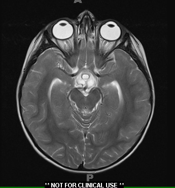
Washington University Experience | NEOPLASM (SELLAR) | Craniopharyngioma, adamantinomatous | 18A3 Craniopharyngioma, Adamantinomatous (Case 18) T2 1 - Copy - Copy
Magnetic resonance imaging in August 2014 shows a 3.3 x 2.8 x 1.8 cm cystic suprasellar mass with an inferior solid enhancing component, and early obstructive hydrocephalus, secondary to compression of the third ventricle. Operative procedure: Endoscopic resection.

