Table of Contents
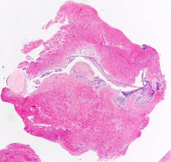
Washington University Experience | NEOPLASM (SELLAR) | Craniopharyngioma, adamantinomatous | 18B1 Craniopharyngioma, Adamantinomatous (Case 18) 20X 2
Sections of the suprasellar lesion show several small fragments of brain parenchyma with marked piloid gliosis, involved in multiple areas by a neoplasm. The neoplasm is characterized by epithelial elements with basal columnar palisades, stellate reticulum, and focally mineralized deposits of 'wet' keratin.

