Table of Contents
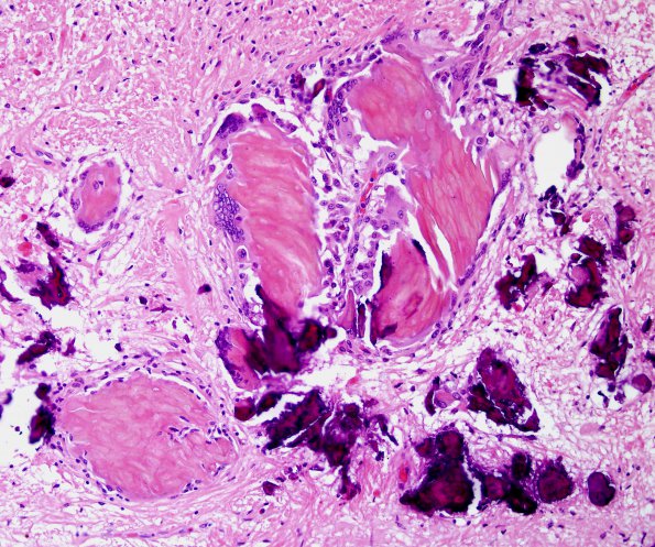
Washington University Experience | NEOPLASM (SELLAR) | Craniopharyngioma, adamantinomatous | 1E3 Cranio & radiation Rx 3 yr prior Case 1) N12 H&E 4.jpg
1E3-5 Higher magnification images of the walls of the third ventricle showing very little residual neoplasm; rather, there are effects of radiation treatment (hyalinized vessels, rarified neuropil), perivascular and scattered foamy macrophages, robust piloid gliosis, and focal lymphocytic infiltrates. (H&E)

