Table of Contents
Washington University Experience | NEOPLASM (SELLAR) | Craniopharyngioma, adamantinomatous | 20A3 Craniopharyngioma (Case 20) 5 copy
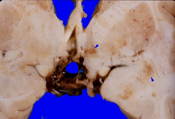
Higher magnification of residual tumor encasing the optic tract shown in image #20A2. Focal discolorations (arrowheads) represent areas of infarction including right orbital gyrus, hypothalamus, optic chiasm and right temporal pole. Soft yellowish, hemorrhagic tumor extended from the level of the anterior commissure and optic chiasm posteriorly to the level of the mammillary bodies. A central cavity, apparently representing remnants of the third ventricle, remained.

