Table of Contents
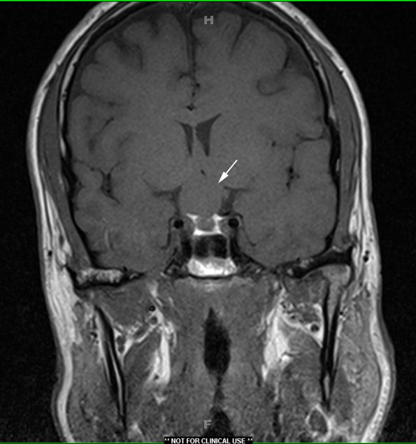
Washington University Experience | NEOPLASM (SELLAR) | Craniopharyngioma, papillary | 1A1 Craniopharyngioma, papillary (Case 1) T1 1 - Copy copy
Case 1 History ---- The patient is a 43-year-old man who recently developed blurry vision, left temporal visual field cut and retro-orbital headache. MRI showed a 2.1 cm cystic suprasellar lesion with mass effect on the optic chiasm and peripheral enhancement. The pituitary gland was relatively normal. Radiographically, craniopharyngioma was favored over macroadenoma. Operative procedure: Right subfrontal craniotomy for tumor excision. ---- 1A1,2 MRI images The neoplasm is seen as an isointense suprasellar mass in T1-weighted non-contrast enhanced (1A1, arrow) and as a hyperintense homogeneous mass in T2-weighted contrast administered scans (1A2).

