Table of Contents
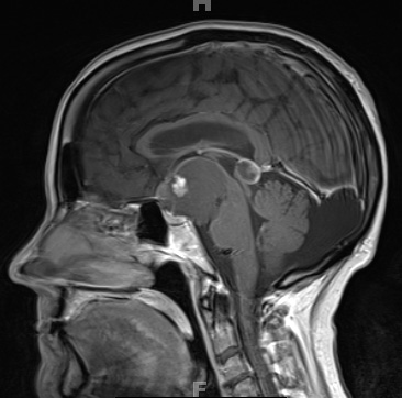
Washington University Experience | NEOPLASM (SELLAR) | Craniopharyngioma, papillary | 2A5 Craniopharyngioma, Papillary (Case 2) T1 W 4 - Copy
The T1-weighted hypointense suprasellar 5.3 x 4.6 x 4.0 cm mass has a 1.1 x 1.0 x 1.2 cm anterior mural nodule which enhances with contrast. The mass is hypertense on a T2-weighted scan (2A6). The mass extends into the prepontine cistern and the interpeduncular cistern as well as into the right Sylvian fissure, and displaces the optic chiasm superiorly as well as the basilar artery laterally. It is seen encasing the bilateral optic nerves, the right A1 segment and the proximal right middle cerebral artery.

