Table of Contents
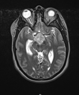
Washington University Experience | NEOPLASM (SELLAR) | Craniopharyngioma, papillary | 3A1 Craniopharyngioma, papillary (Case 3) T2 - Copy
Case 3 History ---- The patient is a 29-year-old woman with a two year history of amenorrhea, 40 pound unintended weight gain, hot flashes, polydipsia, and polyuria. Magnetic resonance imaging showed a 3.1 x 2.9 x 2 cm enhancing mass in the hypothalamic region; the pituitary gland is unremarkable. Operative procedure: Stealth-guided right frontal craniotomy for tumor resection. ---- Magnetic resonance imaging shows a 3.1 x 2.9 x 2 cm T2-weighted variegated MRI hyperintense mass in the hypothalamic region.

