Table of Contents
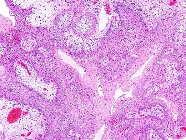
Washington University Experience | NEOPLASM (SELLAR) | Craniopharyngioma, papillary | 3B3 Craniopharyngioma, papillary (Case 3) H&E 10.jpg
Hematoxylin and eosin stained sections of the hypothalamic mass show a neoplasm composed of monomorphic, well-differentiated, non-keratinizing, squamous epithelium supported by broad, loose, pale, hyalinized fibrovascular cores with some chronic inflammation. No maturation of the squamous epithelium, "wet" keratin, or calcifications are seen.

