Table of Contents
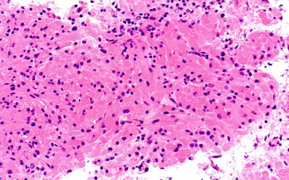
Washington University Experience | NEOPLASM (SELLAR) | TTF-1-positive sellar tumor - Granular cell Tumor | 3A1 Granular cell tumor, infundibulum (Case 3) H&E 1
Case 3 History ---- The patient was a 74-year-old woman with a history of breast cancer. Radiological imaging performed for cancer monitoring revealed a sellar and suprasellar lesion. Visual field testing demonstrated bitemporal superior quadrant defects consistent with a mass effect on the optic chiasm. MRI on 4/13 showed a 1.3 x 1.5 x 2 cm enhancing sellar lesion that was inseparable from the pituitary gland, abutted the cisternal pre-chiasmatic optic nerves, and displaced the optic chiasm superiorly. The radiographic appearance was thought most likely a macroadenoma. The operative procedure performed was an endoscopic endonasal trans-sphenoidal hypophysectomy for tumor resection. ---- 3A1-4 Multiple images of the tumor discovered in this patient composed of large numbers of granule filled tumor cells with significant inflammatory infiltrating cells (H&E)

