Table of Contents
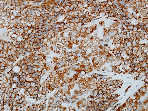
Washington University Experience | NEOPLASM (SELLAR) | Pituitary Adenoma - Pituitary Neuroendocrine Neoplasm | Corticotroph | 14D3 PA, CR in nl (Case 14) YN 5.jpg
Expanded acini (arrows) are distinguished from adjacent tumor in this synaptophysin immunohistochemistry. ---- Not shown: An additional immunohistochemistry panel shows the tumor cells are negative for prolactin, FSH, ACTH, GH, TSH, and LH. p53 stain highlights rare tumor cells. Ki-67 proliferation index highlights 2.1% of tumor cells. ---- Comment: Reticulin stain shows the loss of normal acinar pattern. The morphology and immunoprofile is that of a pituitary adenoma.

