Table of Contents
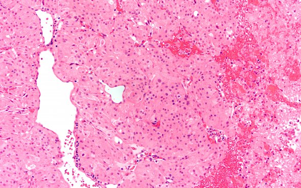
Washington University Experience | NEOPLASM (SELLAR) | Pituitary Adenoma - Pituitary Neuroendocrine Neoplasm | Corticotroph | 18A Pituitary adenoma (Case 18) H&E 1
Case 18 History ---- [53yo male with hypogonadism and acute persistent headache. MRI ~3.7 cm solid and cystic mass likely adenoma expanding the sella, invading the right cavernous sinus, abutting the left cavernous sinus, and showing mass effect on the optic chiasm. Labs: normal levels of GH, Insulin-like Growth Factor, and TSH; reduced levels of PRL, LH, FSH, free T4; and mildly elevated ACTH (85.4 [nl 7.0 to 63]), with normal cortisol. Operative procedure: Endoscopic transsphenoidal hypophysotomy for tumor resection, with intraoperative MRI.] ---- 18A There are relatively large, pleomorphic epithelioid cells with generous eosinophilic cytoplasm, one or more medium-to-large atypical oval (occasionally bizarre) nuclei, and prominent nucleoli. These cells appear in sheets with some weak polarization around delicate blood vessels. In focal discohesive areas the tumor tissue appears papillary. Broad regions of necrosis are evident, with focal perivascular sparing. Mitotic figures are generally common, and focally numerous, reaching a density of 8/10 HPF in one area. No Crooke’s change is identified.

