Table of Contents
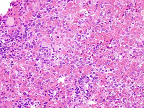
Washington University Experience | NEOPLASM (SELLAR) | Pituitary Adenoma - Pituitary Neuroendocrine Neoplasm | Corticotroph | 20A1 PA CR (Case 20) H&E 1.jpg
Case 20 History ---- [27yo female with hypertension and Cushing's disease. MRI showed a 1.4 cm, post-contrast enhancing sellar mass. Operative procedure: Transsphenoidal endoscopic resection of pituitary tumor.] ---- 20A1-6 The resection specimen shows fragments of adenohypophysis, some which are involved by adenoma. The adenoma has loss of normal acinar architecture (confirmed by a reticulin stain). The tumor cells have an epithelioid appearance with distinct cell borders and abundant amphophilic to basophilic cytoplasm. The tumor nuclei are oval to round with finely stippled chromatin. Rare endocrine atypia is seen. Mitotic figures are not readily identified. There is no necrosis. The uninvolved adenohypophysis shows scattered epithelial acinar cells with Crooke's hyaline change which is highlighted by a CAM5.2 immunostain.

