Table of Contents
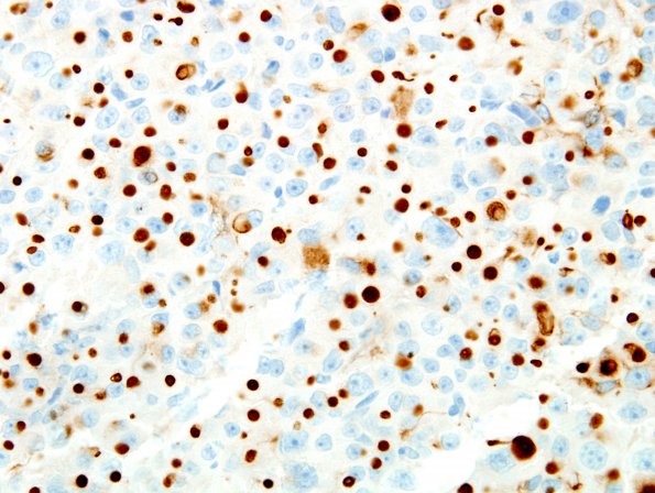
Washington University Experience | NEOPLASM (SELLAR) | Pituitary Adenoma - Pituitary Neuroendocrine Neoplasm | Somatotroph | 5C3 PA (Case 5) CAM5.2 7
The cytokeratin CAM 5.2 shows ball-like cytoplasmic staining in a majority of the tumor cells, consistent with fibrous bodies (rounded aggregates of keratin in growth hormone producing cells). ---- Not shown: A subpopulation of cells also express prolactin. The luteinizing hormone (LH) and follicle stimulating hormone (FSH) stains show scattered positive tumor cells. Adrenocorticotrophic hormone (ACTH) highlights the adjacent normal pituitary gland and rare tumor cells. The tumor cells are nonreactive for TSH. The MIB-1 (Ki-67) proliferative index is low to moderate, reaching 3.2% focally. ---- The histomorphological features and immunostaining profile are consistent with a pituitary adenoma that includes growth hormone production, the latter explaining the acromegaly encountered clinically.

