Table of Contents
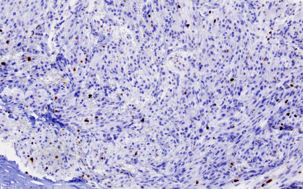
Washington University Experience | NEOPLASM (SELLAR) | TTF-1-positive sellar tumor - Spindle cell oncocytoma (SCO) | 2C Spindle cell Oncocytoma (Case 2) Ki67 1
A minimally increased proliferative index is noted (Ki67 IHC) ---- Not shown: There is focal immunoreactivity for epithelial membrane antigen (EMA), though most of the tumor cells are negative. . A stain for reticulin reveals minimal deposition within the tumor. As in prior specimens, the tumor is strongly immunoreactive for vimentin This latter stain also highlights scattered plasma cells within the tumor. No convincing GFAP expression is found. The immunostains performed on the 1991 specimen show similar features and in addition demonstrate strong immunoreactivity for neuron specific enolase (NSE). Ultrastructural studies on the original specimen demonstrated numerous swollen mitochondria. No definite desmosomes were identified. Apparently, similar features were seen on electron microscopy from the current 2002 specimen. ---- The morphologic, immunohistochemical, and ultrastructural features of this neoplasm are highly unusual and do not fit with either a pituitary adenoma or primary glial neoplasm. Instead, they suggest the diagnosis of "spindle cell oncocytoma". The current case was associated with recurrence after 11 years and this is similarly consistent with a benign or slow growing neoplasm.

