Table of Contents
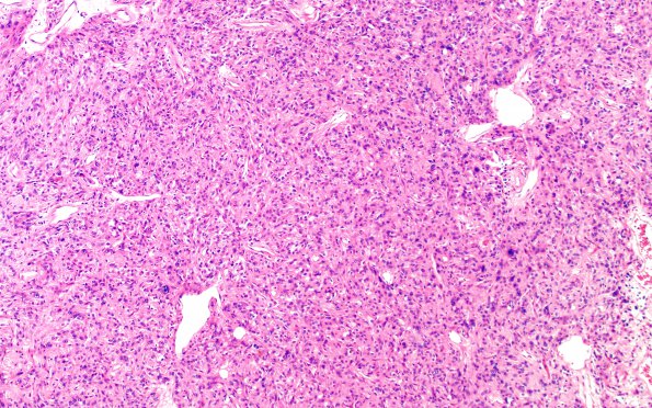
Washington University Experience | NEOPLASM (SELLAR) | TTF-1-positive sellar tumor - Spindle cell oncocytoma (SCO) | 5A3 Spindle cell oncocytoma-pituicytoma DDX (Case 5) H&E 8
5A3-7 Sections of the brain lesion stained with hematoxylin and eosin staining show a neoplasm with variable morphologies. In some areas, the tumor cells are more spindle-shaped and arranged in short fascicles in a storiform pattern. In other areas, the cells are more epithelioid with more abundant cytoplasm and nuclei with more prominent nucleoli. Mitotic figures are rare (1/10HPF). There is no necrosis or vascular proliferation.

