Table of Contents
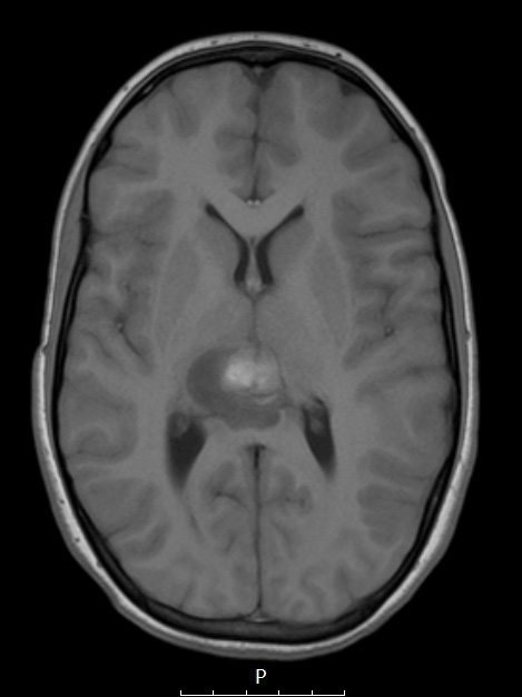
Washington University Experience | NEOPLASMS (EMBRYONAL) | ATRT - Atypical Teratoid Rhabdoid Tumor | 1A1 ATRT (Case 1) T1 no C - Copy
Case 1 History ---- The patient was a 10-year-old girl who presented to the emergency department with complaints of increased confusion, decreased responsiveness, and possible seizures. Operative procedure: Left temporo-parietal craniotomy for excision of temporal lesion using stealth navigation. ---- 1A1-3 On MRI, this lesion shows patchy hyperintensities in the posterior thalamic and adjacent regions in a T1-weighted image (1A1) which enhances with the administration of contrast (1A2,3). The sagittal lesion (1A3) shows continuity of the tumor with the leptomeninges, likely reflecting subarachnoid spread. Multiple foci are seen in the posterior thalamus (3.2 cm), cerebellum (1.5 cm), and left parieto-temporal (2.6 cm) lobe. In addition, there is leptomeningeal enhancement throughout the spinal cord.

