Table of Contents
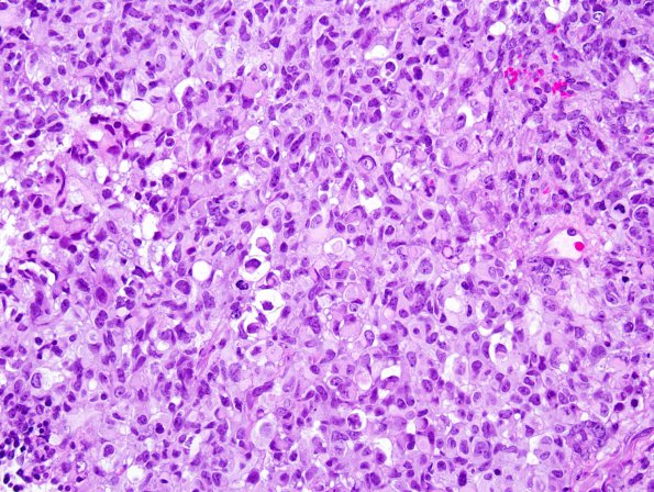
Washington University Experience | NEOPLASMS (EMBRYONAL) | ATRT - Atypical Teratoid Rhabdoid Tumor | 4B3 ATRT (Case 4) H&E 4.jpg
4B3-5 Sections of the right CP angle tumor reveal a highly cellular, relatively solid appearing neoplasm with multiple histologic patterns. In some areas, the tumor resembles a small round cell tumor with primitive-appearing cells containing minimal cytoplasm and a high mitotic index. Other cells appear rhabdoid or gemistocytic, containing eccentric bellies of eosinophilic cytoplasm and occasional globular cytoplasmic inclusions. The tumor cells are mostly arranged in sheets and papillary or pseudopapillary configurations.

