Table of Contents
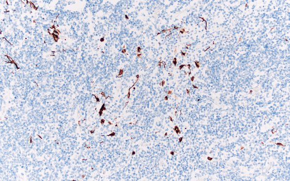
Washington University Experience | NEOPLASMS (EMBRYONAL) | ATRT - Atypical Teratoid Rhabdoid Tumor | 9E2 (Case 9) GFAP 20X 1
Sections which include cerebral cortex show patchy neoplastic infiltration of the parenchyma including areas adjacent to the ependyma and admixed with non-neoplastic germinal matrix. GFAP immunoreactivity shows prominent staining in reduplicated ependyma which likely reflects collapse of the hydrocephalic expanded gliotic ventricular lining

