Table of Contents
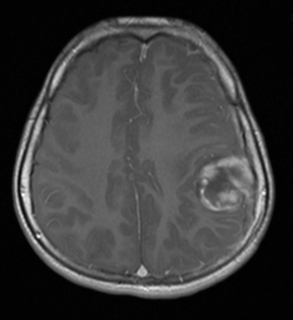
Washington University Experience | NEOPLASMS (EMBRYONAL) | ETMR - Embryonal Tumor Multilayered Rosettes | 5A1 ETANTR (Case 5) T1W 1 - Copy
Case 5 History ---- The patient is a 13-year-old previously healthy male who presented acutely with seizures and altered mental status. MRI on 10/5 shows a 3.4 x 2.2 x 3.1 cm heterogeneously T2-hyperintense left parietal mass centered over the left postcentral gyrus. The mass has irregular peripheral enhancement, no diffusion restriction, and central blood products, with associated mass effect on the central sulcus and precentral gyrus. MRI of the spine shows no evidence of metastasis. Radiological differential includes: Ganglioglioma vs DNET vs metastasis. Operative procedure: Left-sided craniotomy for resection of brain tumor. ---- 5A1,2 MRI shows a mild rim enhancement pattern on a T-1 weighted image with contrast (5A1) and hyperintensity with T2 weighting (5A2)

