Table of Contents
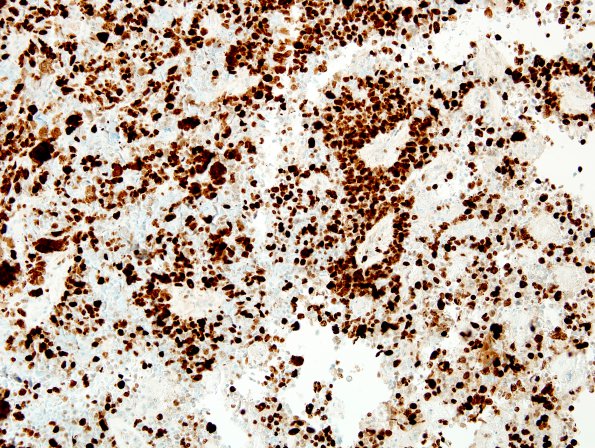
Washington University Experience | NEOPLASMS (EMBRYONAL) | ETMR - Embryonal Tumor Multilayered Rosettes | 6D ETANTR, medulloepithelioma (Case 6) Ki67 1.jpg
The proliferative activity is intense (~70-80%) in rosettes and tumor cells percolating through the neuropil. (Ki67 IHC) ---- Not shown: A neuron-specific enolase immunostain (NSE) shows a similar pattern of staining to synaptophysin. A BAF-47 stain (INI-1) shows retained expression in all tumor nuclei and other resident cells. A glial fibrillary acidic protein immunostain (GFAP) shows only adjacent glial tissues, and rare, entrapped elements, but no definitive glial differentiation within the tumor itself. A neurofilament (NF) stain highlights interspersed neuropil, predominantly at the edges of sheets of tumor, but focally, within tumor as well. A beta-catenin immunostain demonstrates cytoplasmic immunoreactivity exclusively. A reticulin stain does not show a significantly increased reticulin content, but does highlight numerous blood vessels. ---- Comment: In light of the patient's tumor cell previous resections and current specimen this tumor is best characterized as an embryonal tumor. Tests for amplification of the specific miRNA locus on chromosome 19 were performed at St Jude of this material and an earlier resection and the specific amplification of the miRNA cluster on chromosome 19 was not found in either case. Additionally, per report, amplification of the C-MYC or N-MYC loci were not detected in the re-resection material. Additional FISH studies were negative for isochromosome 17q, monosomy of chromosome 6, loss of PTCH-1 (9q), and amplification of C-MYC and N-MYC. The histomorphologic appearance of both tumors contains elements of the previously described entity, ETANTR, but each tumor individually lacks the full complement of diagnostic elements. Among 97 tumor samples of “embryonal tumor with abundant neuropil and true rosettes (ETANTR), ependymoblastoma, and medulloepithelioma analyzed by FISH for amplification of the 19q13.42 locus, four lacked amplification (PMID: 24337497). Rather, these same four displayed polysomy of chromosome 19. As this patient's tumor histopathologically fits this proposed single clinicopathological entity, a lack of amplification may still be consistent with inclusion in this group.

