Table of Contents
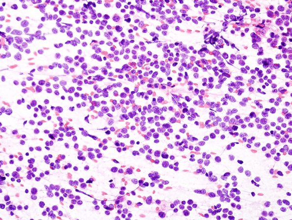
Washington University Experience | NEOPLASMS (EMBRYONAL) | Medulloblastoma, Histologically Defined | Classic Subtype | 20A Medulloblastoma (Case 20) smear.jpg
Case 20 History ---- [10yo male with four weeks of headache, nausea, and vomiting. MRI showed a 3.7 x 3.3 x 4.1 cm heterogeneous T2 iso- to hyperintense and T1 hypointense mass distending the fourth ventricle and extending inferiorly and laterally into the foramen of Luschka. Operative procedure: Craniotomy for posterior fossa tumor resection.] ---- 20A An intraoperative smear shows a hypercellular, patternless proliferation of small cells with small to medium sized hyperchromatic nuclei and minimal cytoplasm. Mitotic figures are numerous, and individual apoptotic cells are readily identified. Overall, the tumor shows a relatively monotonous appearance without any distinctive foci of differentiation or nodularity. Scattered large cells are demonstrated.

