Table of Contents
Washington University Experience | NEOPLASMS (EMBRYONAL) | Medulloblastoma, Histologically Defined | Classic Subtype | 6 A1 Medulloblastoma (Case 6) 5
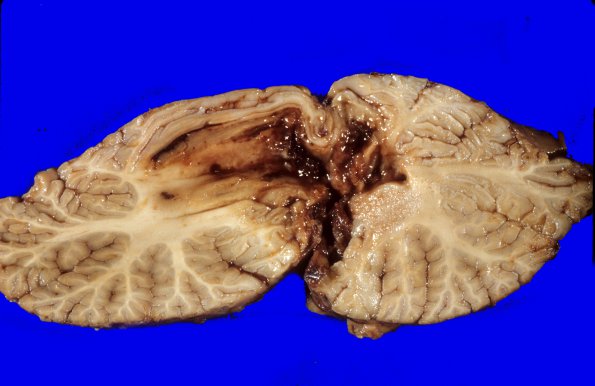
There is a 1½ x 5 x 4 cm hemorrhagic defect in the cerebellar vermis and right cerebellar hemisphere. Coronal sections through the cerebral hemispheres show a large amount of intraventricular hemorrhage involving the left lateral and to a lesser degree the right lateral ventricles. Cross sections through the cerebellum and brainstem show a hemorrhagic cystic necrotic mass involving the midline cerebellar vermis extending into the right and left cerebellar hemispheres and extending anterior into the midbrain tegmentum. Occasional secondary brainstem hemorrhages are noted in the midbrain.

