Table of Contents
Washington University Experience | NEOPLASMS (EMBRYONAL) | Medulloblastoma, Histologically Defined | Desmoplastic-Nodular | 1A1 Medulloblastoma, Desmoplastic (Case 1) 1
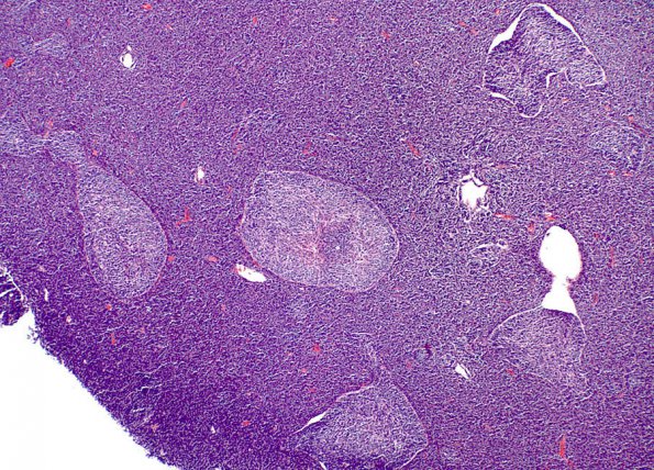
Case 1 History ---- [22yo male with a posterior fossa mass. Clinical diagnosis: Medulloblastoma versus astrocytic neoplasm. Operative procedure: Craniotomy for lesion resection posterior fossa.] ---- 1A1-3 In this case the majority of the specimen had only scattered nodules which made it possible to compare the constituents of individual pale islands. Sections of the posterior fossa tumor show sheets of malignant small round blue cells with hyperchromatic, uniform appearing nuclei and scant cytoplasm. In addition, there are scattered nodules of cells with small round mature appearing nuclei and increased amounts of neuropil in the background. Mitoses are easily identified in the internodular constituents. There is focal necrosis. The tumor extends into the adjacent cerebellar parenchyma along the Virchow-Robin space, a phenomenon designated tumor re-entry from the subarachnoid space. (H&E)

