Table of Contents
Washington University Experience | NEOPLASMS (EMBRYONAL) | Medulloblastoma, Histologically Defined | Ganglion Cell Differentiation | 1A2 Medullo (origl tumor, Case 1) H&E 1
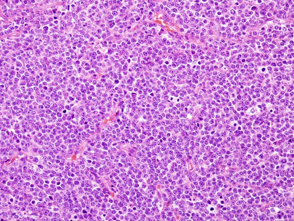
1A2-5 A higher magnification representative section from the original resection specimen reveals a highly cellular small "blue cell" tumor composed of small round to oval nuclei with mild to moderate hyperchromasia and minimal cytoplasm with scattered distinct nodules or pale islands. There are numerous mitotic figures and pyknotic nuclei. Within the pale nodules the tumor cells appear smaller and more mature (neurocyte-like with abundant neuropil in between cells). Occasional Homer Wright rosettes are also seen. Per outside report, the internodular regions were reticulin rich, while there was minimal reticulin deposition within the nodules. (H&E)

