Table of Contents
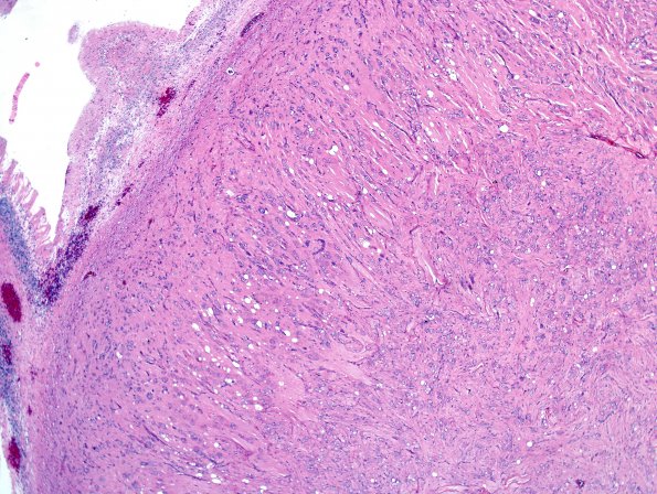
Washington University Experience | NEOPLASMS (EMBRYONAL) | Medulloblastoma, Histologically Defined | Ganglion Cell Differentiation | 2A5 MB differentiation (Case 1) H&E 8
2A5-8 The tumor is much less cellular and is composed predominantly of large mature-appearing ganglion cells with variable dysmorphic features, including clumped Nissl substance and occasional binucleation. In many cases ganglion cells have multiple cytoplasmic vacuoles with a mulberry appearance. In some areas, astrocytic cells are also evident, displaying thin hyperchromatic nuclei with long eosinophilic cytoplasmic processes. Mitotic figures are hard to find and there is no evidence of tumor necrosis. Many of the blood vessels are hyalinized. There are small collections of loose tissue with macrophages and clumps of dystrophic calcification, suggesting the possibility of treatment effect. A fragment of compressed cerebellar tissue is seen at the periphery of the tumor.

