Table of Contents
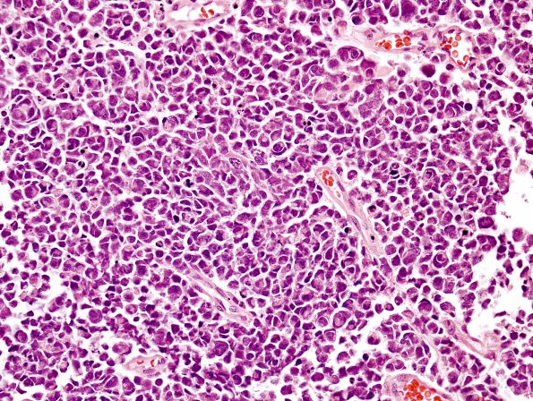
Washington University Experience | NEOPLASMS (EMBRYONAL) | Medulloblastoma, Histologically Defined | Large cell-Anaplastic (LCA) | 4A1 Medulloblastoma, LCA (Case 4) H&E 7
Case 4 History ---- [3yo female with a mass in the fourth ventricle, involving the vermis.] ---- 4A1,2 Histological sections of the posterior fossa mass show a markedly hypercellular neoplasm with an infiltrative growth pattern (evidenced by entrapped neurons and neuropil) and widespread geographic necrosis. The tumor cells are mostly small, with a high nuclear-to-cytoplasmic ratio, usually hyperchromatic nuclei, and scant eosinophilic cytoplasm. Focally, however, the nuclei are considerably enlarged with prominent nucleoli and occasionally eccentrically located cytoplasm, imparting an epithelioid to rhabdoid appearance. However, the majority of the tumor cells appear polygonal, secondary to nuclear molding, with some areas exhibiting 'cell wrapping,' occasionally creating several concentric layers. Elsewhere, the tumor shows small and larger nucleus-free and necrosis-free, vesiculated areas filled by pale, eosinophilic/amphophilic, fine, fibrillar cell processes surrounded by tumor cell nuclei, suggestive of true rosettes.

