Table of Contents
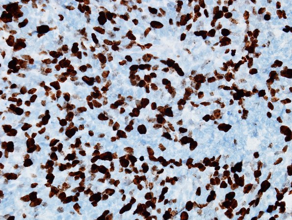
Washington University Experience | NEOPLASMS (EMBRYONAL) | Medulloblastoma, Histologically Defined | Large cell-Anaplastic (LCA) | 8C Medulloblastoma, LCA (Case 8) Ki67.jpg
The Ki67 proliferation index marks an elevated number of tumor nuclei, found in approximately 30% of the total tumor. ---- Not shown: Immunohistochemistry shows that the tumor cells have positive cytoplasmic and paranuclear dot-like reactivity for synaptophysin. Neurofilament is negative in the tumor cells and it highlights the largely solid growth pattern of the tumor, with some minor areas of infiltration. Integrase interactor 1 (INI-1/Baf47/SMARCB1) staining is retained in the tumor nuclei. Beta-catenin is positive in the tumor cell cytoplasm, and not in the nuclei. ---- Comment: The overall histomorphologic and immunophenotypic findings are that of a Medulloblastoma with anaplastic/large cell features, WHO Grade IV.
FISH studies revealed evidence of polysomies (chromosomal gains) of chromosomes 2, 8, and 17 as well as a relative gain of HER2, which may represent presence of isochromosome 17q. There is no evidence for C-MYC or N-MYC amplifications.

