Table of Contents
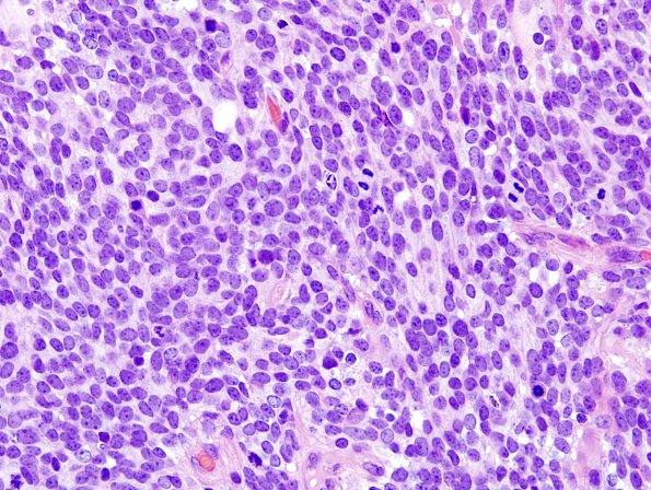
Washington University Experience | NEOPLASMS (EMBRYONAL) | Medulloblastoma, Histologically Defined | Medullomyoblastoma | 1A1 MedMyoB Glial (Case 1) H&E 1.jpg
Case 1 History ---- [The patient is a nine-year-old male with a posterior fossa mass. Operative procedure: Craniotomy for excision of mass] ---- 1A1-4 Sections show a cellular neoplasm consisting of several histologic patterns. ---- 1A1,2 The majority of the specimen consists of a highly cellular, patternless proliferation of cells with small to medium sized hyperchromatic nuclei with minimal cytoplasm. Mitotic activity is frequent and is accompanied by individual cell necrosis with karyorrhectic debris. Tumor cells generally have small nucleoli and focal molding but are not compelling for a large cell/anaplastic tumor type.

