Table of Contents
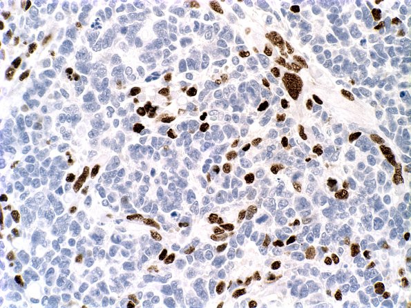
Washington University Experience | NEOPLASMS (EMBRYONAL) | Medulloblastoma, Histologically Defined | Medullomyoblastoma | 3F Medullomyoblastoma (Case 3) Myogenin 2
Foci of rhabdomyoblastic differentiation are confirmed with nuclear staining for myogenin. ---- . Not shown: A stain for epithelial membrane antigen (EMA) is negative. A stain for neurofilament protein highlights axonal formation in the tumor. A histochemical stain for reticulin highlights the desmoplastic regions in the tumor. The morphologic and immunohistochemical features are consistent with a diagnosis of large cell/anaplastic medullomyoblastoma with desmoplastic features.
As a research tool, dual color fluorescence in situ hybridization (FISH) was performed utilizing probes against CEP17, NF1, CEP8, c-myc, BCR and NF2. Polysomies (ie. chromosomal gains) were noted with all of these probes and suggest a state of aneuploidy. Although not entirely specific, this finding is consistent with the degree of anaplasia identified in the tumor (Leonard et al PMID 11453402).

