Table of Contents
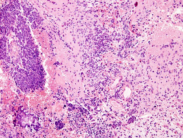
Washington University Experience | NEOPLASMS (EMBRYONAL) | Medulloblastoma, Histologically Defined | Medullomyoblastoma | 4A1 Medullomyoblastoma (Case 4) H&E 1A
Case 4 History ---- [3yo male with a fourth ventricular midline tumor.] ---- 4A1-3 Sections show a medullomyoblastoma. The tumor has two different patterns. In some areas it is composed of diffuse sheets of cells with high N/C ratio, that have oblong nuclei with fine chromatin pattern and show high mitotic activity and individual cell necrosis. These cells show strong membranous positivity for Leu 7, granular and punctate cytoplasmic positivity for synaptophysin and focal paranuclear dot-like cytoplasmic positivity for vimentin. Heavy areas of calcifications are seen focally. Rare individual GFAP mature astrocytes are seen scattered throughout the small neoplastic cells They are negative for cytokeratin, S-100, neurofilaments, actin, desmin, and myoglobin. The morphology and immunophenotype of this area is consistent with a medulloblastoma

