Table of Contents
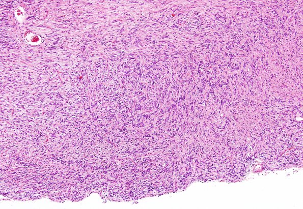
Washington University Experience | NEOPLASMS (GLIAL) | Angiocentric glioma | 2B4 Angiocentric Glioma (Case 2) H&E 5
2B4-6 The tumor tissue shown in these images is more densely cellular but still consists of a monomorphic population of bipolar tumor cells with elongate oval, hyperchromatic nuclei, small nucleoli, eosinophilic cytoplasm, and generally indistinct cytoplasmic borders. These cells appear over a microcystic and piloid background of tumor cell processes, and exhibit several architectural patterns, including perivascular pseudorosette formation involving both large and small caliber vessels, and widespread 'spongioblastic' nuclear palisading. (H&E)

