Table of Contents
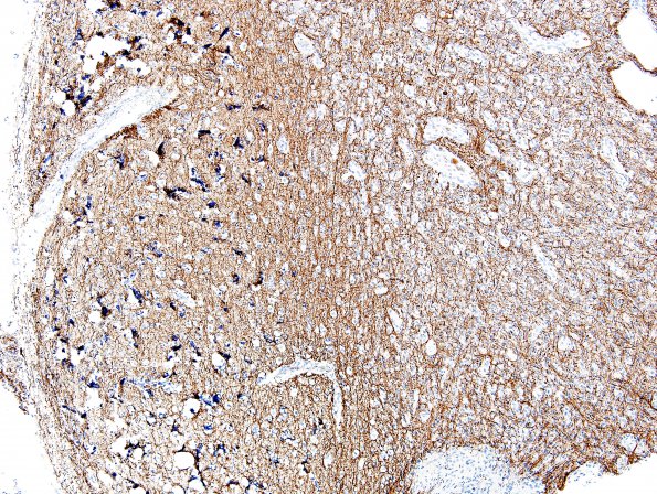
Washington University Experience | NEOPLASMS (GLIAL) | Angiocentric glioma | 2D1 Angiocentric Glioma (Case 2) NF 5
2D1-3 Deeper layers of cortical neurons are entrapped within the tumor, but show no evidence of dysmorphic features, evidencing the tumor's infiltrative growth pattern. The adjacent superficial cortical layers show reactive changes, including concentric microcalcifications, punctate mineralization of small caliber vessels, and scant hemosiderin deposition, accompanied by only occasional tumor cells. Mitotic figures are not observed. No ganglion cells, eosinophilic granular bodies (EGB), or Rosenthal fibers are identified. Reactivity for neurofilament protein (NF) highlights axonal processes within the tumor parenchyma that range from sparse to abundant; each of these stains reflect the tumor's infiltrative growth pattern. ---- Other IHC Stains: Reactivity for synaptophysin showed a geographic pattern similar to that of neurofilament protein, with dense granular staining of the neuropil of the uninvolved cortex and slightly less intense staining of the infiltrated deeper layers of cortex. Reactivity for proliferation marker Ki67 is 2.3%.

