Table of Contents
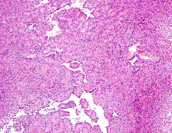
Washington University Experience | NEOPLASMS (GLIAL) | Astroblastoma | 1A2 Astroblastoma, WHO II (Case 1) H&E 12.jpg
1A2-8 There is extensive hyalinization in the background and mild nuclear pleomorphism. The majority of tumor cells contain oval nuclei with delicate chromatin and clear cytoplasm. Although there are multiple foci of coagulative necrosis, mitotic figures are infrequent and there is no definite microvascular proliferation. Some areas also show intersecting fascicles of spindled tumor cells. Focally, the tumor cells are arranged around central blood vessels with a perivascular nuclear free zone. However, the tumor cells in the vague perivascular pseudorosettes are predominantly cuboidal with broad processes, rather than delicate fibrillary processes.

