Table of Contents
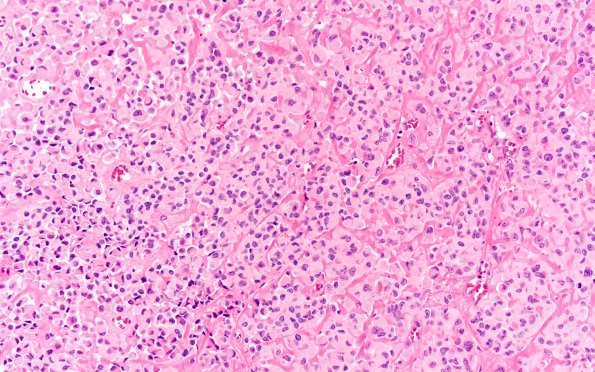
Washington University Experience | NEOPLASMS (GLIAL) | Astroblastoma | 2A1 Astroblastoma, recurrent (Case 2) H&E 1
Case 2 History ---- The patient is a 16 year old girl with several resections for an astroblastoma. Histologic materials shown represent the most recent resections. ---- 2A1-4 Hematoxylin and eosin stained sections of the right frontoparietal biopsy material show a hypercellular, dura based neoplasm with relatively sharp demarcation from the surrounding brain parenchyma. Most of the cells present a sheeted architecture. The vessels are often hyalinized. The tumor cells are pleomorphic. Many of them have eccentric, hyperchromatic nuclei and abundant glassy, eosinophilic cytoplasm, resembling gemistocytes. Some tumor cells have nuclear pseudoinclusions. Multinucleated cells are also present. Mitotic activity is brisk (up to five mitoses per high power field). There's focal hemosiderin deposition. Endothelial hyperplasia is present focally. No necrosis is identified.

