Table of Contents
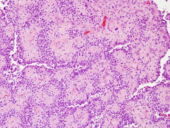
Washington University Experience | NEOPLASMS (GLIAL) | Astroblastoma | 3A3 Astroblastoma, focal anaplasia (Case 3) H&E area A 20X.jpg
Sections reveal a solid-appearing moderately cellular neoplasm arranged in sheets and papillary configurations. The blood vessels are markedly hyalinized. The tumor cells are moderately pleomorphic and consist predominantly of epithelioid to spindled cells with moderate quantities of clear to eosinophilic cytoplasm. In some areas the tumor cells are arranged radially around central blood vessels. The cells surrounding the central blood vessels appear cuboidal to columnar with broad processes radiating towards the vessels. Mitotic figures are rare. There are scattered multi-nucleated giant cells with bizarre atypical nuclei, the latter most likely representing degenerative changes. In most areas, the tumor has a low grade appearance. However, focal microvascular proliferation and necrosis is evident.

