Table of Contents
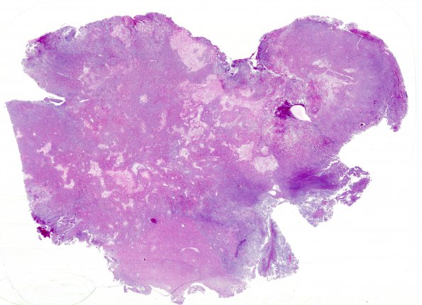
Washington University Experience | NEOPLASMS (GLIAL) | Astroblastoma | 4A1 Astroblastoma, focal anaplasia (Case 4) 1 H&E WM
Case 4 History ---- The patient is a six year old girl with a partially cystic, enhancing left fronto-parietal neoplasm ---- 4A1-3 Sections reveal a markedly hyalinized glial neoplasm. The neoplasm displays a pushing border with the adjacent brain parenchyma. The neoplasm is composed of medium to large cells with glial to epithelioid cytology. Throughout most of the neoplasm, the cells are uniform and round to polygonal with oval nuclei harboring a delicate chromatin pattern and eccentric eosinophilic cytoplasm, resembling gemistocytic or rhabdoid cells. In these regions, there is extensive perivascular hyalinization, low to moderate cellularity, mild pleomorphism, and rare mitotic activity. Focally, a compact cellular architecture is evident with minimal perivascular hyalinization, increased pleomorphism, and increased mitotic activity. Atypical mitoses are also identified. In some regions, a papillary architecture is evident. Perivascular pseudorosettes are numerous. Unlike the classic ependymal pseudorosette, these are characterized by cuboidal, epithelioid cells with broad foot processes radiating towards the vascular lumen. Small foci of "infarct-like" necrosis are identified without any associated pseudopalisading or hypercellularity. Focal endothelial hyperplasia is evident.

